
Unravelling the Brain’s Eating Regulation: Recent research led sheds light on the brainstem’s pivotal role in controlling appetite and eating habits, revolutionising our understanding of this complex process.
In a new study published in Nature, a team led by Zachary Knight, PhD, from the University of California, San Francisco (UCSF), offers new insights into how our brainstem controls appetite and eating habits. This research not only challenges long-standing theories but also opens up potential avenues for more effective weight-loss treatments.
Neurological Interplay of Taste and Satiety
Recent studies challenge the belief that stomach cues mainly dictate eating. They highlight taste’s crucial role in eating patterns. Researchers emphasize that food taste, not fullness, determines eating speed and quantity.
Key to this discovery are PRLH neurons in the brainstem. These neurons respond instantly to taste perceptions. Their rapid activation differs from the slower gastric signal responses. PRLH neurons act as regulators, modulating food intake based on taste.
This breakthrough reshapes our understanding of appetite control. It shows how taste perception immediately affects brain cells. This insight is vital for understanding brain regulation of food intake. It helps in tackling obesity and eating disorders.
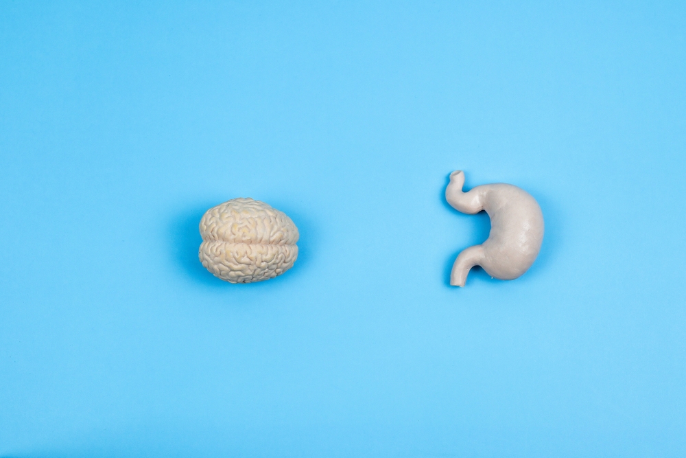
The findings contribute significantly to neurogastroenterology. They reveal the brain-digestive system connection and the role of sensory experiences. This research prompts reconsideration of taste in dietary management and weight control strategies.
Gut Signals and Long-Term Satiety
Complementing the role of taste-focused PRLH neurons, the study by UCSF researchers also sheds light on another crucial player in appetite control: GCG neurons. These neurons represent a different facet of our body’s complex satiety mechanisms, being primarily influenced by mechanical signals from the gut. Unlike the rapid response of PRLH neurons to taste, GCG neurons are attuned to the physical aspects of food consumption, notably the volume and mechanical pressure within the gastrointestinal (GI) tract.
GCG neurons are integral to monitoring and responding to the amount of food consumed, playing a pivotal role in generating long-term feelings of satiety. As food is ingested and the stomach distends, these neurons receive mechanical feedback, which then triggers their activity. This process is crucial for the brain’s ability to assess how much food has been eaten and to signal when enough has been consumed, providing a sense of fullness that persists well after the meal has ended.
One of the most intriguing aspects of GCG neurons is their role in the release of GLP-1 (Glucagon-like peptide-1), a hormone known for its appetite-suppressing properties. GLP-1 is a key target for several contemporary weight-loss medications, including the widely discussed drug Ozempic. The UCSF study’s insights into how GCG neurons function and how they interact with GLP-1 offer valuable clues into the mechanisms of these drugs. Understanding this interaction better could lead to the development of more effective treatments for obesity and related metabolic disorders.
Technological Breakthrough in Brainstem Imaging
The groundbreaking research conducted by the UCSF team was made possible by significant technological advancements in brainstem imaging, spearheaded by Truong Ly, PhD, a graduate student in Zachary Knight’s lab.
“We’ve uncovered a logic the brainstem uses to control how fast and how much we eat, using two different kinds of signals, one coming from the mouth, and one coming much later from the gut,” said Knight. “This discovery gives us a new framework for understanding how we control our eating.”
These innovative imaging techniques represent a leap forward in neuroscience research, particularly in studying the brainstem, an area that has historically been challenging to observe due to its deep location within the brain.
Ly’s development enabled real-time brainstem observation in awake mice, previously impossible. This method accurately shows brain function during normal behavior, like eating. Monitoring the active brainstem is vital for understanding brain functions. It provides insights into brain behavior during eating.
This technology allowed the team to see how taste affects PRLH and GCG neurons. This observation revealed the neurons’ roles in taste and eating. This breakthrough revives interest in Ivan Pavlov’s digestion theories. Pavlov emphasised sensory experiences in digestion, a previously underexplored area. The new imaging method opens research possibilities in neurogastroenterology and neuroscience. It allows detailed study of deep-brain structures.
Implications for Weight-Loss Strategies
Understanding the dual role of taste and gut signals in appetite control could lead to more personalised and effective weight-loss strategies. The team plans to further explore how these signals interact during a meal, potentially offering new insights into appetite suppression.
References
- From the First Bite, Our Sense of Taste Helps Pace Our Eating | UC San Francisco. (2023, November 22). From the First Bite, Our Sense of Taste Helps Pace Our Eating | UC San Francisco. https://www.ucsf.edu/news/2023/11/426631/first-bite-our-sense-taste-helps-pace-our-eating
- Ly, T., Oh, J. Y., Sivakumar, N., Shehata, S., La Santa Medina, N., Huang, H., Liu, Z., Fang, W., Barnes, C., Dundar, N., Jarvie, B. C., Ravi, A., Barnhill, O., Li, C., Lee, G., Choi, J., Jang, H., & Knight, Z. A. (2023, November 22). Sequential appetite suppression by oral and visceral feedback to the brainstem. Nature. https://doi.org/10.1038/s41586-023-06758-2


