

While mammograms remain the cornerstone of breast cancer screening, breast ultrasound offers valuable additional insight when doctors need a closer look at lumps, dense breast tissue, or unclear findings.
Breast cancer is one of the leading causes of death among women globally. About 670,000 individuals succumbed to it in 2022, according to the World Health Organization (WHO).


Early diagnosis is key to managing this disease. While mammograms take center stage in discussions about breast cancer screening, an advanced ultrasound system plays a vital supporting role when diagnostic accuracy is questioned.
Taking control of your health starts with understanding your options. Here’s a straightforward look at what a breast ultrasound is and how it works, and why your doctor might recommend one.
How A Breast Ultrasound Works
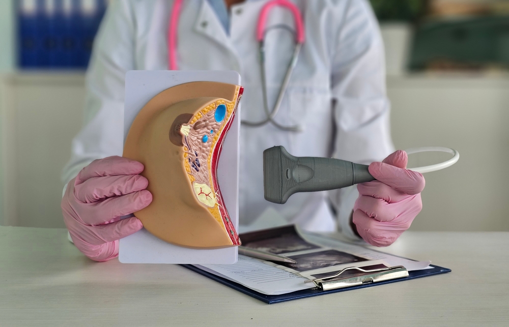
Breast ultrasound is a diagnostic imaging test that uses high-frequency sound waves to create detailed images of your breasts. This clinical procedure doesn’t involve radiation, unlike X-rays or mammograms. That said, it’s exceptionally safe for everyone, including pregnant women and breastfeeding mothers.
During the procedure, a trained medical professional, called a sonographer, applies a clear ultrasound gel to your skin. They will gently move the wand over your breast tissue. The transducer sends sound waves into your body, which bounce off your internal tissues and echo back to the machine. A computer translates those echoes into real-time visual images on a monitor.
As the technology is versatile, high-quality ultrasound equipment for OB-GYN clinics and dedicated imaging centers has become an absolute cornerstone of women’s healthcare. With these tools, providers can quickly evaluate patients’ concerns without compromising accuracy.
How It Differs From a Mammogram
As the world becomes more serious about breast health, many find themselves performing self-exams or undergoing routine cancer screening. Some might wonder why they need an ultrasound if they’re updated with their annual mammogram schedules. To answer this question, you have to understand that they use different technologies and scan your breast tissues in different ways.
Mammograms use low-dose X-rays to take a two-dimensional image of the breast. They’re the gold standard for regular screening because they excel at spotting tiny calcium deposits, which can be an early sign of cancer. However, mammographic imaging struggles to distinguish between a tumor and glandular tissue.
Ultrasounds, meanwhile, provide a dynamic and real-time look at the tissue from multiple angles, making it ideal for identifying different types of breast lesions. An ultrasound technician can target a specific spot of concern and see beneath the surface layers without needing to compress the chest.
These two are complementary. A sonography is rarely used as a standalone routine screening tool. Even so, it’s a powerful device that can investigate and provide solid answers that a mammogram can’t answer.
Why You Might Need One
Doctors will order a breast sonography for a variety of diagnostic reasons, but mainly for enhancing their assessment capacities.
Below are the most common scenarios that require one:
- Investigating a lump: Your doctor may have felt a hard spot or thickening during a physical breast exam. The procedure enables the medical team to see what’s causing it.
- Clarifying an inconclusive mammogram result: Breast implants can sometimes obscure results. Specialists use ultrasound as a follow-up test to get a clearer and more detailed view.
- Differentiating a solid mass and a cyst: A tissue lump and a cyst (a fluid-filled sac that’s usually non-cancerous) can both show up as white spots on a mammogram. Ultrasound images show the difference between them.
- Checking symptoms in certain populations: Younger women have dense breast tissue, while pregnant and nursing women should avoid unnecessary radiation exposure. An ultrasound is the safest choice when patients under this demographic complain of breast pain or any unusual changes.
If a breast sonography reveals a suspicious solid mass rather than a cyst, the doctor may use a specialized biopsy needle to gently extract a tiny breast tissue sample for additional clinical procedures.
Recent figures show that 985,400 cases of breast cancer were reported in Asia in 2022. The disease took the lives of 315,100 individuals in the region during the same period. High incidences of mortality were reported among older women in progressive countries.
However, specialists are encouraging early diagnostic tests, as younger women may unknowingly develop it. For instance, a study has discovered that ethnic Chinese women are more likely than Australians to develop breast cancer at an early age, likely due to diet and lifestyle. Dr. Stephen Birell, an Australian oncologist, has noticed such a discrepancy in his practice, telling abc.net.au, “They get it at a young(er) age than in the West.”
Breast Ultrasound vs. MRI
Breast MRI (magnetic resonance imaging) is another option, particularly for screening women with a high genetic risk of breast cancer or checking the integrity of breast implants. This diagnostic tool taps magnets and radio waves to create cross-sectional images.
While capable of delivering vivid imaging results, it’s incredibly sensitive and can sometimes flag harmless breast lumps as suspicious. This could lead to unnecessary anxiety and follow-up biopsies. A whole breast ultrasound is generally better at correctly identifying healthy tissues and preventing false alarms.
Advances in ultrasound technology are enhancing diagnostic accuracy and treatment planning. A 3D breast ultrasound system, also called the Advanced Automated Breast Ultrasound (ABUS), can perform automated image quality assessment. This feature guides technicians on the proper positioning, while algorithms analyze the image quality simultaneously.
Final Thoughts
Staying on top of your breast health can feel less overwhelming if you know your options and how they work. An ultrasound schedule isn’t necessarily a cause for alarm. In many cases, it’s a reassuring step that empowers your healthcare providers to keep you healthy. Most scans turn out normal, but if your doctor finds something that needs further attention, just know that early detection and proactive protection give you an advantage.