


Repairing a leaking heart valve without opening the chest is one of cardiology’s most complex procedures. At St. Antonius Hospital in the Netherlands, Medical Channel Asia saw how Philips is using AI-powered image guidance to help clinicians navigate a beating heart. This brings together imaging, precision and teamwork to improve patient care.
A beating heart is one of the most difficult places for a doctor to work.


Inside the operating room at St. Antonius Hospital in the Netherlands, a team of cardiologists, echocardiographers, radiographers and nurses stand around a patient. Their eyes are fixed not on the heart itself, but on multiple imaging screens. Every movement of the catheter is guided by live X-ray and ultrasound. Meanwhile, every decision is made while the heart continues beating.
Success is measured in millimetres.
It is here that Philips believes artificial intelligence (AI) can make one of medicine’s most technically demanding procedures safer, more intuitive and more precise.
Medical Channel Asia visited Philips’ Innovation Campus in Eindhoven and St. Antonius Hospital to see how the company’s AI-powered image guidance technology is supporting minimally invasive heart valve repair. This is a procedure that could offer hope to patients who are too frail for conventional open-heart surgery.
What Happens When a Heart Valve Leaks?

At the centre of the procedure is the mitral valve, one of four valves responsible for keeping blood flowing in the right direction through the heart.
When the valve closes properly, oxygen-rich blood is pumped efficiently to the rest of the body. But in people with mitral valve regurgitation, the valve no longer seals completely. Instead, blood leaks backwards every time the heart contracts.
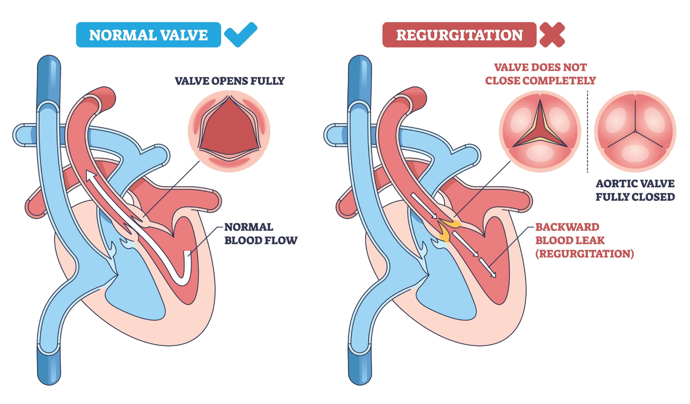
The consequences can be serious.
Patients often develop breathlessness, dizziness, fatigue, swelling of the legs and, over time, heart failure. Around one in ten people over the age of 75 develop problems affecting either the mitral or tricuspid valve, yet many remain untreated because they are not suitable candidates for conventional surgery.
Treating Mitral Valve Regurgitation
For many years, severe mitral regurgitation was primarily treated through open-heart surgery. While highly effective, the operation requires opening the chest, temporarily stopping the heart and placing the patient on a heart-lung machine while surgeons repair or replace the damaged valve.
Not every patient is well enough to undergo such a major operation.
Older adults, patients with weakened heart function and those living with multiple medical conditions often face a much higher surgical risk. For these patients, minimally invasive transcatheter valve repair has opened up an important alternative.
Instead of opening the chest, doctors insert a catheter through a vein in the groin and carefully guide a small repair implant into the beating heart, where it is used to bring the leaking valve leaflets back together.
The approach is considerably less invasive and can shorten recovery, but it introduces a new challenge.
Without directly seeing the heart, clinicians must navigate delicate instruments inside a constantly moving organ using only medical imaging.
Seeing What Doctors Cannot
Unlike open-heart surgery, where surgeons have a direct view of the valve, transcatheter repair relies entirely on imaging.
Throughout the procedure, the heart continues beating naturally. Rather than being connected to a heart-lung machine, the patient remains on a beating heart. Meanwhile, the team guides the repair device using fluoroscopy (live X-ray) and transoesophageal echocardiography (TEE).
Working on a beating heart makes the procedure technically demanding, but it also offers an important advantage.
Because blood continues flowing through the valve, clinicians can immediately assess whether the repair has successfully reduced the leak. If the device has not been positioned optimally, it can be repositioned before being permanently released.

“The valve is moving all the time,” Dr Martin Swaans explained during a procedural walkthrough. “It’s a beating heart procedure.”
Success depends on much more than simply reaching the valve.
The repair device must approach from exactly the right angle before grasping both valve leaflets. Dr Swaans compared it to buttoning a shirt.
“If you button up your shirt wrong,” he said, “you are going to destroy the valve or actually make it worse.”
Adding to the complexity, clinicians are working inside a three-dimensional structure while much of the information they receive comes from two-dimensional imaging.
Where AI Comes In
Recognising this challenge, Philips has developed an AI-powered image guidance platform designed to support the heart team throughout the procedure.
Rather than making decisions for clinicians, the system automatically identifies the repair device on live fluoroscopy images and detects whether its arms are open or closed. It also predicts its trajectory and integrates this information with ultrasound images. As a result, it provides a clearer understanding of where the device is inside the heart.
Before treatment, clinicians can plan where the implant should ideally be positioned based on the patient’s anatomy. During the intervention itself, the AI continuously helps the team visualise the device as it moves through the beating heart. In addition, it supports communication between the interventional cardiologist, echocardiographer and the rest of the multidisciplinary team.
During a live procedure observed at St. Antonius Hospital, that collaboration was evident throughout the operation.

As the team positioned the repair device, they discussed the amount of residual leakage and assessed whether the implant had grasped the valve leaflets adequately. They also considered whether additional implants would provide a better result. Because the heart continued beating throughout the procedure, they could immediately evaluate how each adjustment affected blood flow. Consequently, they could decide whether to reposition the device or implant another clip.
The result is not an autonomous system replacing clinicians, but an additional layer of information to support clinical judgement.
As structural heart procedures become increasingly complex, Philips says it is working with partners to integrate AI-powered image guidance across more devices and procedures. This is helping more clinicians perform minimally invasive interventions with greater confidence.
A Second Chance at Life



For the team inside the operating room, the discussion centred on millimetres, imaging angles and valve anatomy. For patients, however, the outcome is much simpler – another chance at life.
On 18 June 2011, while playing football with her children, she suddenly collapsed.
She was rushed to St. Antonius Hospital, where doctors discovered she had suffered a spontaneous coronary artery dissection rather than conventional coronary artery disease. She was clinically dead before being placed on a heart-lung machine and undergoing emergency open-heart surgery using an artery taken from her leg.
Although the operation saved her life, her recovery was anything but straightforward.
After spending three days in a coma and two weeks in hospital, she was discharged home. Soon afterwards, fluid accumulated in her body, forcing her back into hospital for another three months. The damage left her heart functioning at only around one-third of its normal capacity. She also developed severe mitral valve regurgitation.
Another open-heart operation was considered too risky.
Hope Through Innovation
At the time, the minimally invasive clip procedure was still in its early stages of clinical adoption. After doctors successfully treated an older patient using the new technique, Erica was offered the same procedure just two weeks later. For someone considered too frail for another open-heart operation, it represented a chance that would not have existed only a few years earlier.
The results transformed her life.
Her valve leakage improved from severe to moderate, allowing her to gradually regain her independence after around a year of rehabilitation. Today, while she no longer runs or participates in endurance sports, she otherwise leads a largely normal life.
Every year on 18 June, she celebrates what she calls her “second birthday”.
She has since travelled with Dr Swaans across the Netherlands, sharing her story and helping educate other patients about minimally invasive heart valve repair.
“It’s so important that these innovations continue. That we keep finding ways to help more patients.” she said, reflecting on how imaging technology and AI continue to evolve. “With today’s imaging technologies and AI, physicians can identify the optimal location much more accurately.”
For Erica, however, technology is only part of the story.
“Carpe Diem. Seize the day.” she said. “None of us knows whether we’ll still be here tomorrow.”
The Future of Structural Heart Care
Artificial intelligence will never replace the expertise required to perform complex heart procedures. Clinicians remain responsible for every decision, from identifying the leaking segment of the valve to determining the optimal position of the implant.
But by helping doctors visualise devices more clearly and integrate multiple imaging modalities, AI is becoming another member of the heart team. It can also help doctors navigate one of the body’s most challenging organs.
For patients like Erica, that progress is measured not in algorithms or software updates, but in another chance to celebrate a birthday.

