

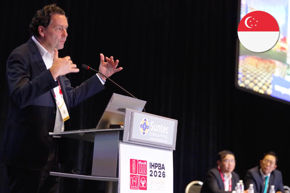
At UltraGreen’s sponsored symposium at IHPBA 2026, surgeons explored how ICG fluorescence can help locate tumours, map liver anatomy and guide resection—and why interpreting the signal is key to its clinical value.
During liver surgery, finding a suspicious area is only the beginning. Surgeons must understand its relationship to surrounding tissue, decide where to divide the liver and assess whether the intended removal is complete. An additional image can help, but its value depends on how it informs those decisions.
Indocyanine green (ICG) fluorescence imaging offers another way to examine the operative field. ICG is a fluorescent dye that binds rapidly to proteins in the blood after injection. When illuminated with near-infrared light, it emits a signal that a dedicated camera captures and displays on a monitor. The liver takes up the dye and excretes it into bile. These properties allow surgeons to visualise blood-flow patterns and biliary structures, depending on when and how ICG is administered, and have applications in identifying lesions, assessing surgical margins and outlining anatomical structures.
These applications formed the focus of “ICG-Assisted Procedures in Liver Surgery”, an UltraGreen-sponsored lunch symposium at IHPBA 2026 in Singapore.
From Fluorescent Signals to Surgical Decisions
“How Green Is Green?” captures a practical challenge in fluorescence-guided surgery: deciding what a visible signal means for the operation. Professor Alexander Vahrmeijer, President of the International Society for Fluorescence Guided Surgery (ISFGS) and head of the Green Light Leiden research group, explained that brightness can vary with camera position. Surgeons therefore need to consider where fluorescence appears and how it relates to the tissue being removed.
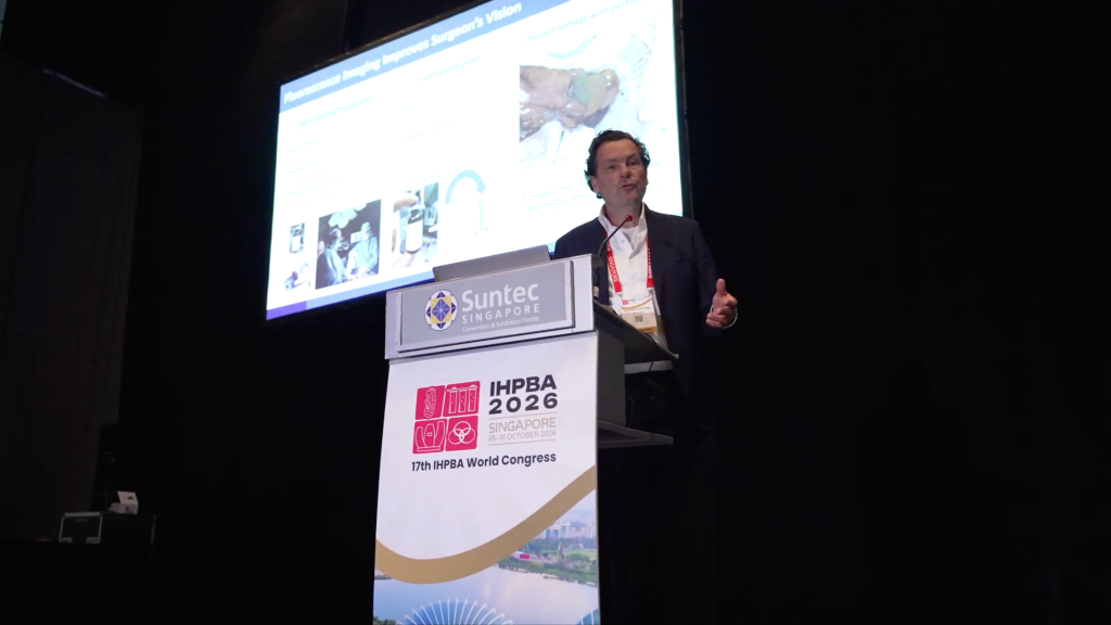
Around colorectal liver metastases, nearby liver cells may clear ICG less efficiently, creating a fluorescent rim around the tumour. During resection, this pattern can provide feedback on how closely the surgeon is approaching the lesion.
Prof Vahrmeijer described checking both the removed specimen and the remaining surgical cavity. Fluorescence at the resection surface can prompt closer assessment and, where appropriate, additional tissue removal.
The 2024 MIMIC study examined this approach across eight Dutch centres, analysing 201 patients with 316 colorectal liver metastases. Additional removal of fluorescent tissue was associated with an increase in margin-negative resections from 87.4% to 92.4%. A negative margin was defined as at least 1 mm between the tumour and the cut surface.
The fluorescence findings frequently prompted intraoperative adjustments. “In one in three patients, the surgeon in the trial adapted their technique a little bit based on the first findings,” Prof Vahrmeijer noted.
These findings support fluorescence as an adjunct to ultrasound and conventional imaging for margin assessment in this setting. The single-arm study did not establish a survival benefit.
Mapping the Liver Before Resection
Beyond assessing tumour margins, fluorescence can help outline the liver territory a surgeon intends to remove. In the prospective HEPATOFLUO study, injecting ICG into a branch of the portal vein allowed the corresponding liver territory to be identified in 16 of 20 patients.
Prof Brian Goh, Head and Senior Consultant at the Department of Hepato-pancreato-biliary and Transplant Surgery, Division of Surgery & Surgical Oncology, Singapore General Hospital and National Cancer Centre Singapore, illustrated a related application through a recording of a central liver resection.
The aim was to remove the central portion while preserving blood supply to the liver on either side. Fluorescence provided a visual check that his team had controlled the intended pedicles serving that territory.
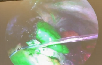
“We give ICG to confirm that we have taken the appropriate pedicles. After confirming we have gotten the correct pedicle, we can complete the resection.” He shared,
“So ICG gives us confidence to perform this kind of surgery precisely.”
A More Complete View During Surgery
Fluorescence and intraoperative ultrasound offer complementary views: fluorescence can reveal superficial lesions, while ultrasound assesses deeper tissue and internal structures. A 2026 meta-analysis of 11 studies involving 558 patients and 894 cancerous liver lesions reported detection sensitivity of approximately 99.8% with both techniques, compared with 86.8% for ultrasound alone.
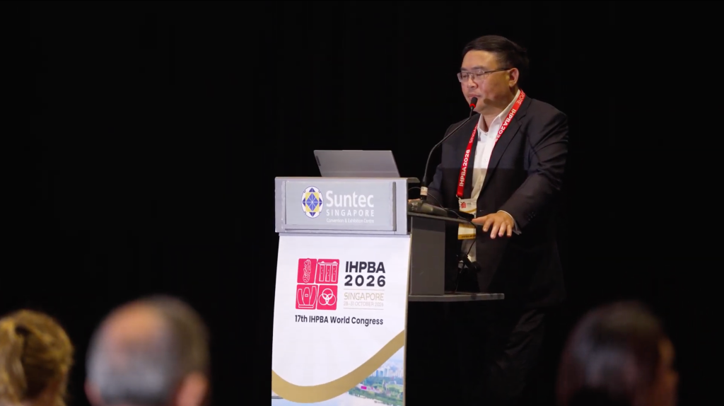
Prof Goh illustrated their complementary use through a case of suspected recurrent cancer after a liver transplant. Scarring and adhesions obscured the lesion, and ultrasound left its location uncertain.
“We did ultrasound, we weren’t sure, and then surprisingly the tumor is actually on the diaphragm.”
Fluorescence helped locate the lesion on the diaphragm rather than within the transplanted liver, changing the planned resection.
“So in this case, we did the laparoscopic resection of the diaphragm instead of the liver and closed it up. Otherwise, it would have been very challenging for us to tell where the tumor is.”
Refining the Signal
Obtaining useful fluorescence images involves more than choosing when to switch on the camera.
Previous treatment can influence the fluorescence surgeons see. A 2025 study found reduced ICG accumulation around colorectal liver metastases in patients who had received chemotherapy, suggesting that their imaging protocols may need adjustment.
The ongoing MIMIC-II study is investigating changes to ICG dose and timing in this group. Its findings could help establish protocols better suited to chemotherapy-treated patients, giving surgeons more consistent information during resection.
This focus on tailoring the technique to the patient extends to interpretation: treatment history and the condition of the underlying liver remain important when assessing the signal.
A Measured Approach to Wider Adoption
Prof Goh described the growing role of fluorescence in minimally invasive practice.
“Indications for this imaging are rapidly expanding, and the use of fluorescent imaging is an essential tool in the armamentarium of anyone doing HPB surgery, especially via the MIS (minimally invasive surgery) approach,” he said.
Effective use requires careful patient selection and interpretation alongside established imaging techniques. Dr Glenn Bonney, a hepatobiliary, pancreatic and transplant surgeon on the Local Organising Committee and Scientific Programme Committee for IHPBA 2026, highlighted the complexity of liver surgery today.
“Liver surgery has advanced so far that the combination of metabolic and chemo-induced liver injury means we are rarely operating on ‘normal’ livers,” he said.
Bonney, a Senior Consultant at PanAsia Surgery Group and Adjunct Associate Professor at the National University of Singapore, acknowledged advances in technology, dosing and detection. However, underlying liver function and biliary stasis affect fluorescence, with false-positive and false-negative findings remaining a challenge.
“It’s incumbent on senior surgeons to remain measured in any approach of technology.”
What This Means for Patients
For patients, the practical question is whether fluorescence could help clarify a specific part of their operation. Asking their surgeon what it may add – and why it is appropriate for their case – can help them understand the surgical plan.
When used thoughtfully, fluorescence offers another way to tailor surgical care to the individual patient, for better patient outcomes.
This article is supported by UltraGreen.

